
Osteoporosis
LEARN MOREOsteoporosis
What is it?
Osteoporosis is a disease in which bone weakening increases the risk of a broken bone. It is the most common reason for a broken bone among the elderly
Bones that commonly break include the vertebrae in the spine, the bones of the forearm, and the hip.
Until a broken bone occurs there are typically no symptoms. Bones may weaken to such a degree that a break may occur with minor stress or spontaneously.
Osteoporosis may be due to lower-than-normal maximum bone mass and greater-than-normal bone loss.
Bone loss increases after menopause due to lower levels of estrogen.
Osteoporosis may also occur due to a number of diseases or treatments, including alcoholism, anorexia, hyperthyroidism, kidney disease, and surgical removal of the ovaries.
Prevention of osteoporosis includes a proper diet during childhood and efforts to avoid medications that increase the rate of bone loss. Efforts to prevent broken bones in those with osteoporosis include a good diet, exercise, and fall prevention.
Lifestyle changes such as stopping smoking and not drinking alcohol may help.
Osteoporosis in women
In women, bone density (or mass) progressively increases up to the age of about 30, when the bones are at their most robust. After this period, the bone density gradually decreases. The rate of bone loss accelerates after menopause.
If the body is unable to maintain an adequate level of bone formation, the bones continue to lose density and become increasingly fragile, eventually resulting in osteoporosis.
Types of osteoporosis
There are two main types of osteoporosis:
- Primary osteoporosis
- Secondary osteoporosis
Primary osteoporosis
More than 95% of cases of osteoporosis in women and 80% of cases in men are due to primary osteoporosis. Most cases occur in postmenopausal women and elderly men.
One of the main causes of osteoporosis is the lack of estrogen, in particular their rapid decrease, which occurs during menopause.
Most men over the age of 50 have higher estrogen levels than postmenopausal women, although they also have a decline in the latter due to aging.
Low estrogen levels are associated with osteoporosis in both men and women. Lack of estrogen increases the incidence of bone depletion and causes rapid bone loss.
In men, the low level of male sex hormones also contributes to osteoporosis. The loss of bone density can be even greater if there is insufficient calcium or vitamin D intake.
Low levels of vitamin D cause a lack of calcium and increase the activity of the parathyroid glands, causing an excessive release of parathyroid hormone, which can in turn promote bone depletion.
Secondary osteoporosis
Less than 5% of female osteoporosis and about 20% of male osteoporosis is secondary.
Examples of diseases that can cause secondary osteoporosis are chronic renal failure and hormonal disorders (especially Cushing’s disease, hyperparathyroidism, hyperthyroidism, hypogonadism, high levels of prolactin and diabetes mellitus).
Some types of cancer, such as multiple myeloma, and other chronic diseases, such as rheumatoid arthritis, can cause secondary osteoporosis.
Some drugs that can cause secondary osteoporosis are, for example, progesterone, corticosteroids, thyroid hormones, some chemotherapy and anti-seizure drugs.
Cigarette smoking, excessive consumption of alcohol or caffeine can also cause or contribute to secondary osteoporosis.
Primary osteoporosis risk factors
- Family members suffering from osteoporosis
- A diet with low calcium and vitamin D intake
- Sedentary life
- White or Asian race
- Slim constitution
- Premature menopause
- Cigarette smoke
- Excessive consumption of alcohol and caffeine
Idiopathic osteoporosis
Idiopathic osteoporosis is a rare form of osteoporosis. The term idiopathic simply means that the cause is not known.
This type of osteoporosis occurs in pre-menopausal women, men under the age of 50, children and young adults who have normal hormone and vitamin D levels and have no obvious reason for bone weakness.
Symptoms
Osteoporosis itself has no symptoms; its main consequence is the increased risk of bone fractures.
Osteoporotic fractures occur in situations where healthy people would not normally break a bone; they are therefore regarded as fragility fractures.
Typical fragility fractures occur in the vertebral column, rib, hip and wrist. they are the most commonly related to osteoporosis. The symptoms of a vertebral collapse (“compression fracture”) are sudden back pain, often with radicular pain (shooting pain due to nerve root compression) and rarely with spinal cord compression or cauda equina syndrome.
Risk factors for osteoporotic fracture can be split between nonmodifiable and (potentially) modifiable. In addition, osteoporosis is a recognized complication of specific diseases and disorders.
Medication use is theoretically modifiable, although in many cases, the use of medication that increases osteoporosis risk may be unavoidable.
Diagnosis
The diagnosis of osteoporosis can be made using conventional radiography and by measuring the bone mineral density.
- All women from 65 years of age
- Depending on the likelihood of an underlying problem, investigations for cancer with metastasis to the bone, multiple myeloma, Cushing’s disease and other above-mentioned causes may be performed.
- All men and women who have had a previous fracture with no or little force, even if the fracture occurred at a young age
- Adults from 65 years of age who suffer from unexplained back pain or who have lost at least 3 centimeters (1.2 inches) in height
- People whose bones appear thin or who have vertebral compression fractures on radiographic examination
- People at risk of developing secondary osteoporosis
Some radiographic findings may indicate osteoporosis, however the diagnosis is confirmed by bone densitometry.
Treatments
- Calcium and Vitamin D.
- Monoclonal antibody medications.
- Hormone-related therapy.
- Bone-building medications
Back pain resulting from a spinal compression fracture should be treated with pain relievers and sometimes with heat and massage and / or supportive devices (such as a brace).
Calcitonin may be prescribed to decrease the pain caused by vertebral fractures. Drug therapy includes the use of bisphosphonates, a class of drugs capable of inhibiting bone resorption. They are also used in the elderly hormonal support therapies, anabolics, vitamin D.
Back-strengthening exercises can help improve chronic back pain. Typically, the subject should avoid bed rest and lifting weights. The subject should do resistance exercises under load.
Fractures due to osteoporosis need to be treated.
Preventio
La prevenzione dell’osteoporosi è in genere più efficace del trattamento, dal momento che è più facile prevenire la perdita di densità ossea che ripristinare la densità una volta perduta. La prevenzione dell’osteoporosi prevede:
- managing risk factors (for example, quitting smoking and avoiding excessive consumption of alcohol and caffeine)
- performing resistance exercises (such as walking, climbing stairs or lifting weights)
- Calcium and Vitamin D.
Most experts recommend about 30 minutes of resistance exercise under load per day. A physical therapist can develop a safe physical activity program and demonstrate how to perform daily activities while minimizing the risk of falls and spinal fractures.
Interestingly, high levels of physical activity in premenopausal women, as in the case of female athletes, can actually cause a small decrease in bone density, because such exercises inhibit the production of estrogen by the ovaries.
Strengthening exercises of the back muscles
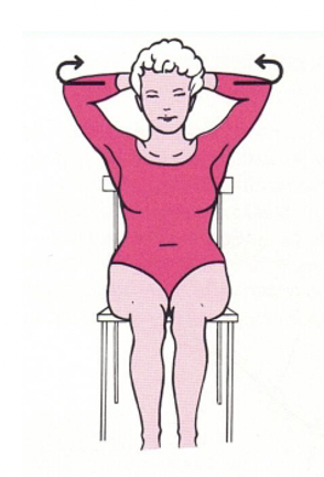
1. Respiratory and dorsal extensor muscles
This exercise favors the relaxation of the pectoral muscles and the extension of the back, with an increase in the tone of the paravertebral muscles. Sitting in the chair, fingers interlaced behind the head. The elbows must be brought back inhaling deeply: on exhalation the flexed arms are brought forward.
Repeat this exercise slowly 20-30 times.
2. Shoulder muscles
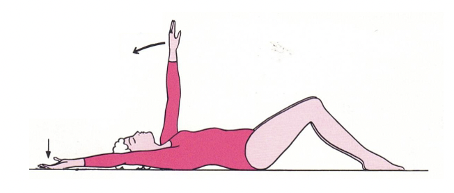
Lie on your back, bend your knees slightly. The arms are extended above the head and alternately pressed against the support surface.
Perform 20 times for each arm.
3. Hip and knee extensors
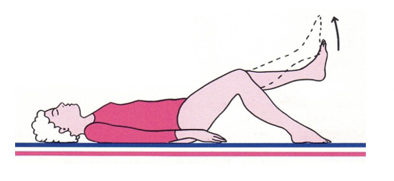
Lying on the support surface with the arms along the sides, the knees are partially bent, resting the sole of the foot on the surface. Alternately, the legs are slowly extended and lifted to a vertical position, then returned to the starting position.
Perform this movement 20 times for each leg.
4. Abdominal and dorsal extensor muscles
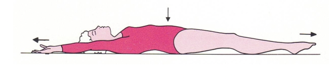
This exercise represents a form of muscle relaxation, useful for improving the tone of the back and abdominal muscles. Lie on your back and bring your arms above your head. In this position, after a deep inhalation, the maximum possible extension of the body is sought, maintaining it for a few seconds.
Repeat slowly 10-15 times.
5. Back extensors
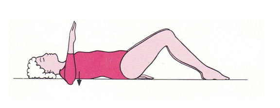
Lie on your back, bend your knees slightly. With the fingertips pointing towards the ceiling, the elbows are flexed to 90° and simultaneously pressed against the support surface.
Repeat slowly 20-30 times.
6. Shoulder muscles
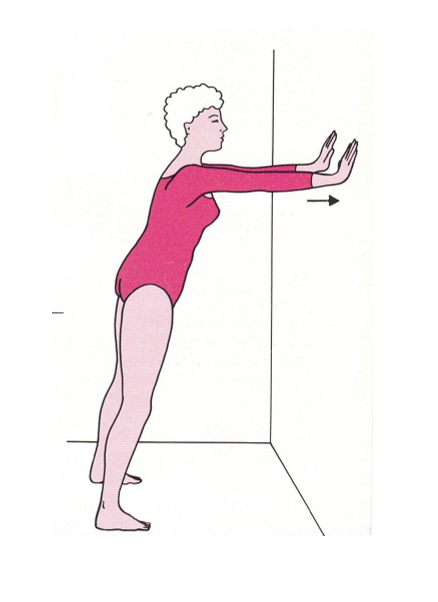
Standing, at a distance of 50-60 cm. from the wall, place the palms of the hands on the wall, at eye level. Inhaling deeply, gradually apply pressure for 2-3 seconds.
Repeat 20-30 times.